Quick links / on this page
Photon biological effects:
Proton biological effects:
- Project 2: Common analysis methods for extracting clinical proton-RBE can lead to spurious conclusions
- Project 3: Developing lineal energy spectrum-based proton RBE models
Introduction
All of my work in computational microdosimetry is done with the idea in mind that an improved understanding of the physical interactions of ionizing radiation can improve our ability to predict what the biological effects of ionizing radiation will be. In this section I report on some projects I have lead or been involved with towards that goal.
Predicting early DNA damage with microdosimetry
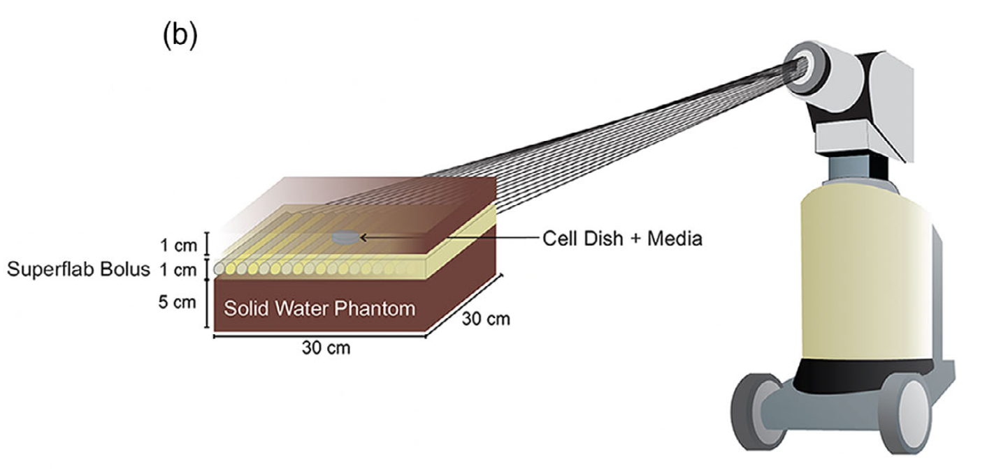
Following my time at McGill University, Dr. Mirta Dumancic began working in Dr. Shirin Enger’s lab as a postdoctoral scholar. She worked on a variety of projects, one of which involved taking the MicroDose Monte Carlo software I had developed and previously reported on and applying it to determine lineal energy spectra in cell lines irradiated with various brachytherapy and megavoltage photon therapy sources. As part of this work she paired with biologists in the Enger lab including Joanna Li and others. For this study HeLa and PC3 cell lines were irradiated with 50 and 250 kVp X-rays, Ir-192, and 6 MV photons from a Varian linear accelerator. Their setup for Ir-192 irradiations is depicted below:
Using cell and nucleus size distributions for HeLa and PC3 retrieved from the literature a pouring simulation using LAMMPS was performed to develop a three dimensional cell model. This cell model was imported in to MicroDose and lineal energy spectra in the cells were calculated for several bracytherapy sources and for megavoltage photons.
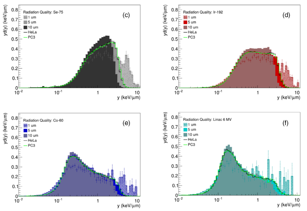
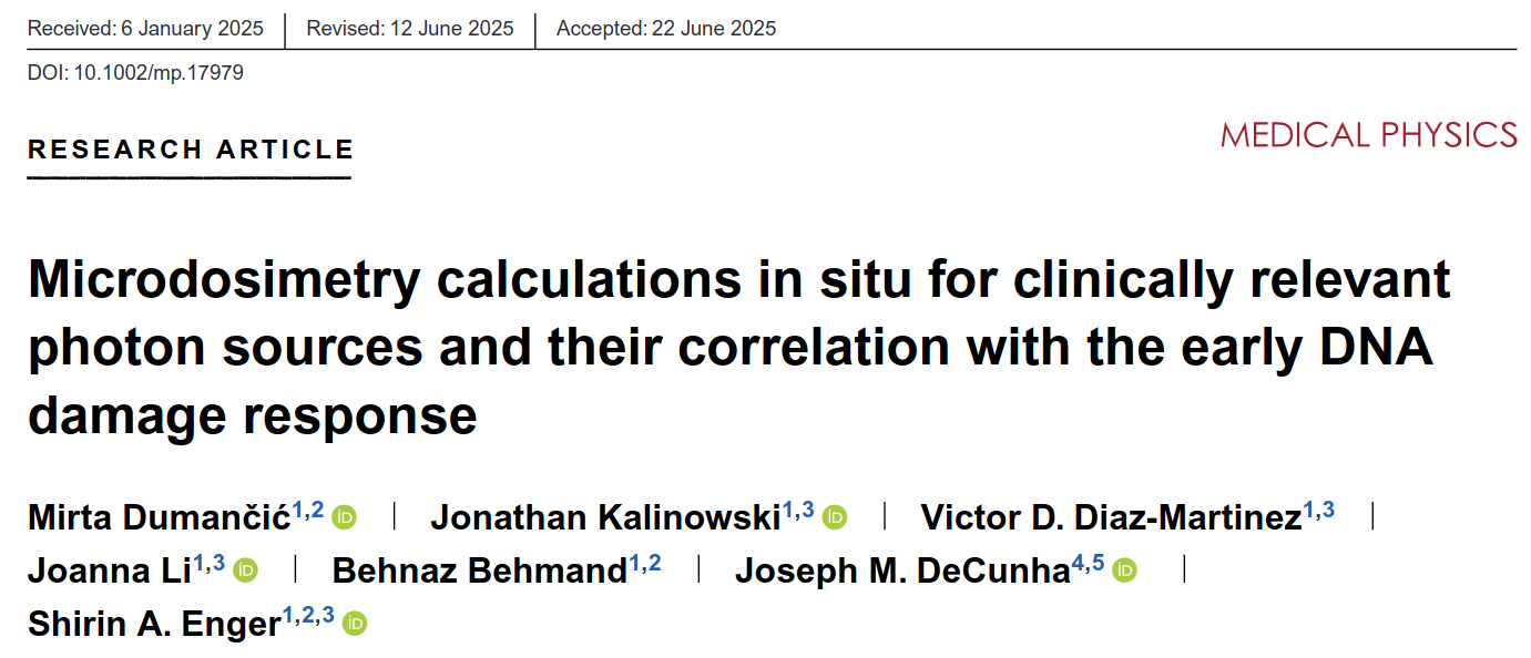
Dose-mean lineal energy (the expectation value of the dose spectrum of lineal energy) was retrieved for 50 and 250 kVp X-rays, Ir-192, and 6 MV photons. It was shown that dose-mean lineal energy was highly correlated with the induction of gamma-H2AX foci (a surrogate for DNA double strand break damage) immediately after radiation. From this, we were able to demonstrate the utility of microdosimetric quantities for predicting DNA damage immediately following irradiation. This work was published in Medical Physics:
Common analysis methods for extracting clinical proton-RBE can lead to spurious conclusions
During my time as a PhD candidate at MD Anderson Cancer Center, I turned my focus from microdosimetry and predictions of the biological effects of new and proposed brachytherapy isotopes, to the microdosometry and biological effects of proton therapy.
Protons and heavy ions are known to possess a quantity described as spatially variable relative biological effectiveness (RBE). Despite more than six decades of knowledge that spatially-variable proton-RBE exists, we still do not make use of proton-RBE in treatment planning of clinical proton cases. This suggests that to some degree (the extent of which is debated), plans developed for proton therapy are suboptimal because we are assuming the biological effects of protons are spatially consistent. In my opinion, the greatest reasons that contribute to our lack of considering proton-RBE during treatment planning are 1.) we don’t know that proton-RBE effects observed in cells are applicable to in-vivo tissue response in humans and 2.) we lack clinical evidence that spatially variable proton-RBE impacts treatment outcomes and tissue complications.
It was not until the 2016 work by Peeler et al. that it was claimed that clinical evidence of spatially-variable proton-RBE effects could be observed. In the Peeler study, a correlation between image enhancement regions on post-treatment MRI images and regions in which proton linear energy transfer (LET) existed was observed.
At MD Anderson, I worked in Radhe Mohan’s lab, the same lab in which Christopher Peeler worked. As part of my interest in proton-RBE, I went back and reanalzyed the cohort in the original Peeler study. I did this work in an attempt to explain why the Peeler study detected a correlation between proton-LET and image change risk and some work from our colleagues at Massachusetts General Hospital failed to find any such correlation. I hypothesized that the analysis method used may be playing a major role in whether such a correlation was found. For this work I applied the four primary analysis methods for this type of study which had been described in the literature.
When I applied an analysis approach known as mixed effects regression, which essentially involves developing a risk model for each individual patient, I found that only 8/14 patients showed evidence of an association between proton-LET and image change risk (below):
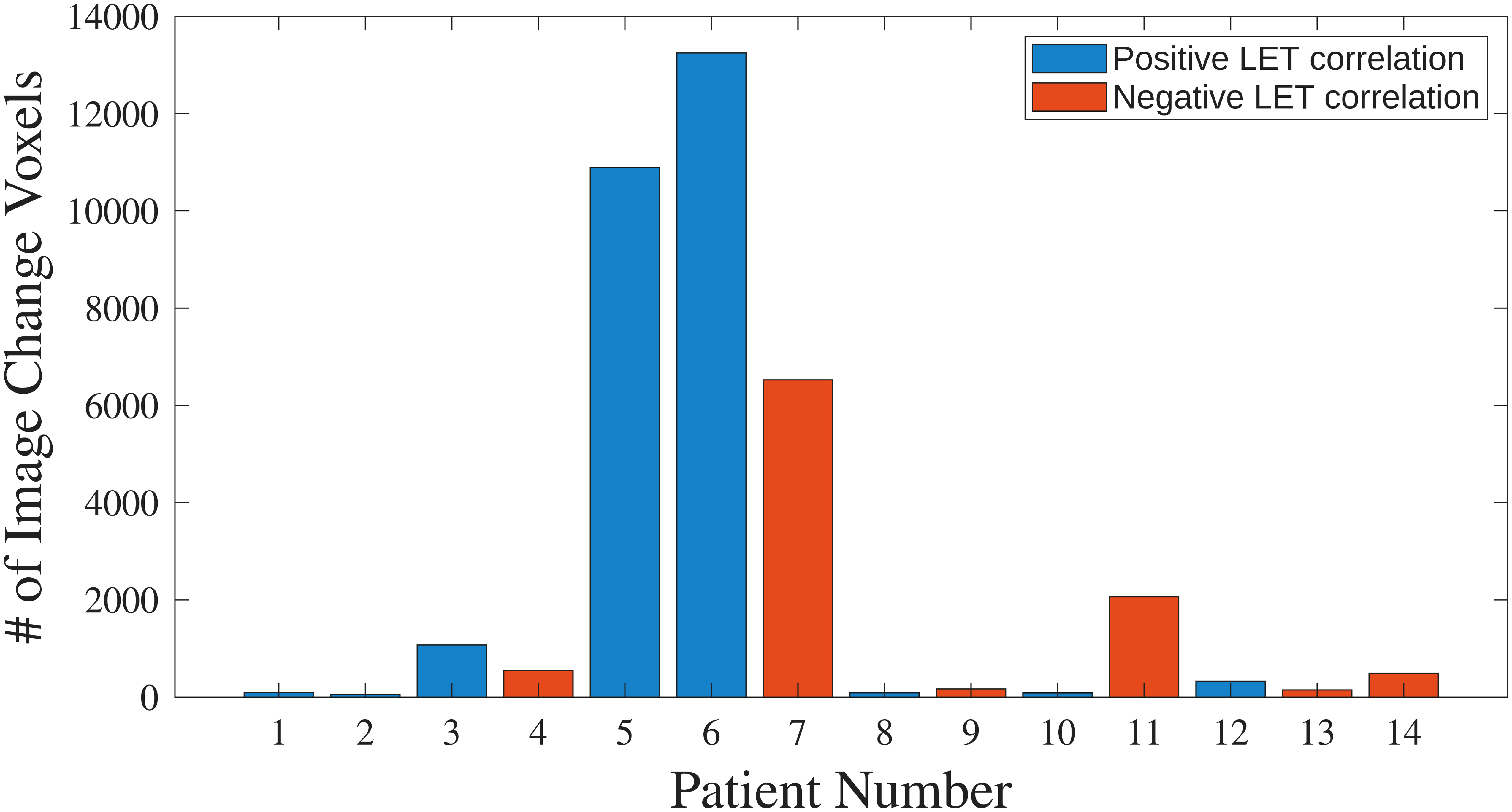
As part of this study, I discovered that a data mishandling practice known as pseudoreplication had been conducted. Pseudoreplication occurs when highly correlated data points, such as many thousands of voxels from a single individuals brain, are erroneously treated as uncorrelated. This mistake was made in the Peeler study and several others like it. Under reanalysis using more appropiate mixed effects method no statistically signifigant relationship between proton-LET and image change risk was found. We published on this work in Advances in Radiation Oncology.
Developing lineal energy spectrum-based proton RBE models
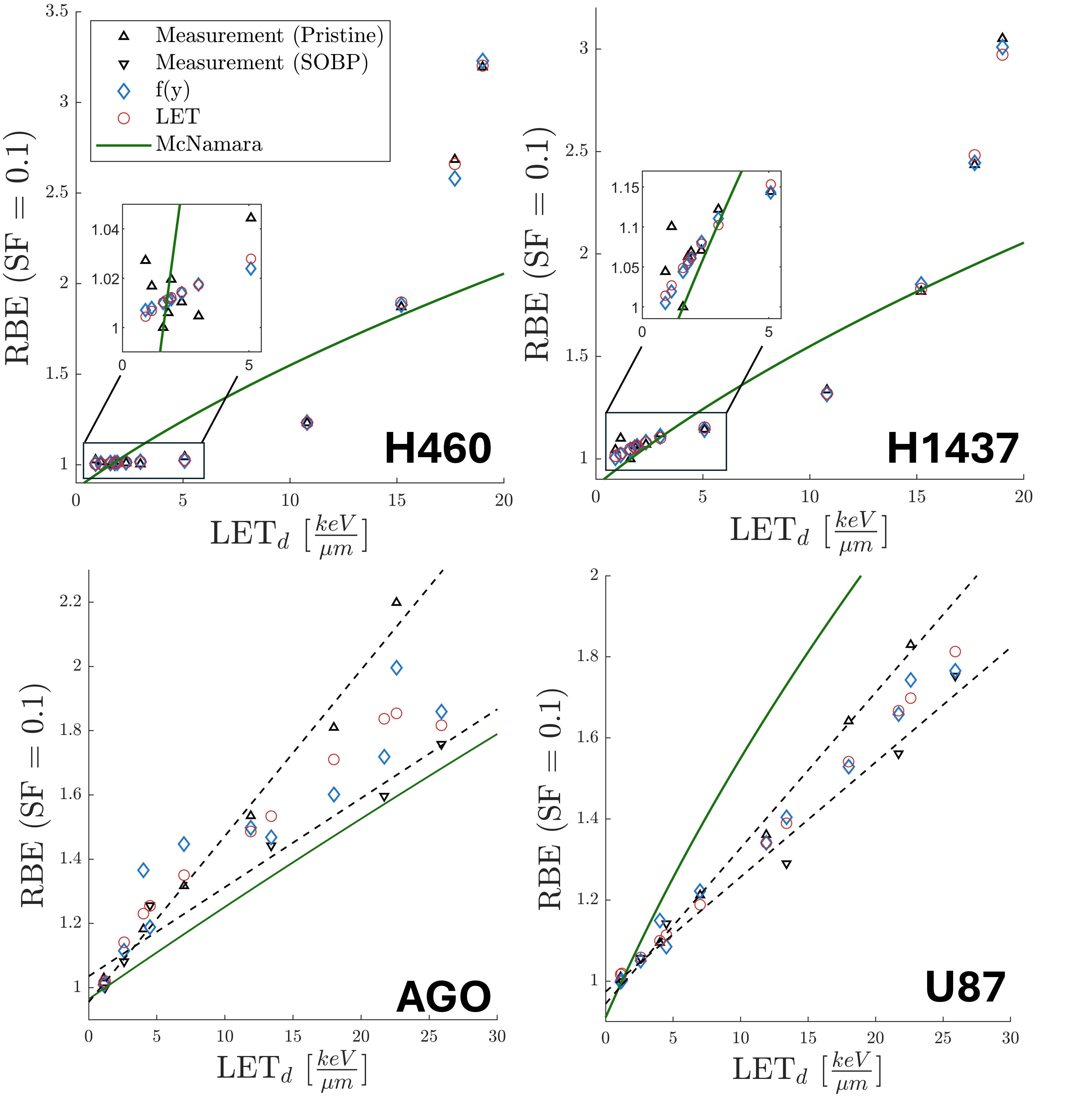
While I have published several studies on the calculation of lineal energy for brachytherapy sources and in proton therapy, I am yet to publish the logical extension of this work; that being, to develop lineal energy spectrum-based biological effects models. I currently have work under review with Precision Radiation Oncology to do exactly that. My work addresses a lack of lineal energy spectrum-based biological effects models which exist. While, some models such as the Local Effect Model (LEM) and Generalized Stochastic Microdosimetric Kinetic Model (GSM) make use of proton energy spectra or lineal energy spectra, these models are either semi-mechanistic, or rather opaque and hard for researchers outside of the groups that developed them to employ (this is especially the case for the LEM). My most recent work addresses this by creating purely analytical lineal energy spectrum-based proton-RBE models. In the figure below it shown that creating cell-line specific RBE models can deliver performance highly favorable over RBE models developed to predict RBE across a wide variety of cell lines: